State of the art and detailed ophthalmologic examination to assess diseases and determine the general health of the eyes with extreme accuracy. Our experts are some of the best around. We hand select our specialists from many fields of ophthalmology, so we are equipped to meet your vision need, whatever it is!
Keeping an Eye on Your Vision
To maintain healthy eyes, start with consistent eye exams. Regular comprehensive eye exams allow us to find, diagnose and treat eye issues before they fully develop. On top of making sure you eyes are healthy, eye exams help us determine the best methods for improving your vision. Young or old, vision is critical to a happy and healthy lifestyle. With specialists in every area, you can trust that the doctors of Gulf Coast Eye Institute will provide you with excellent vision and excellent eye health.
Whether you’re nearsighted or farsighted, have presbyopia, astigmatism or a degenerative eye disease, we will work with you to improve your sight.
Routine Eye Exams
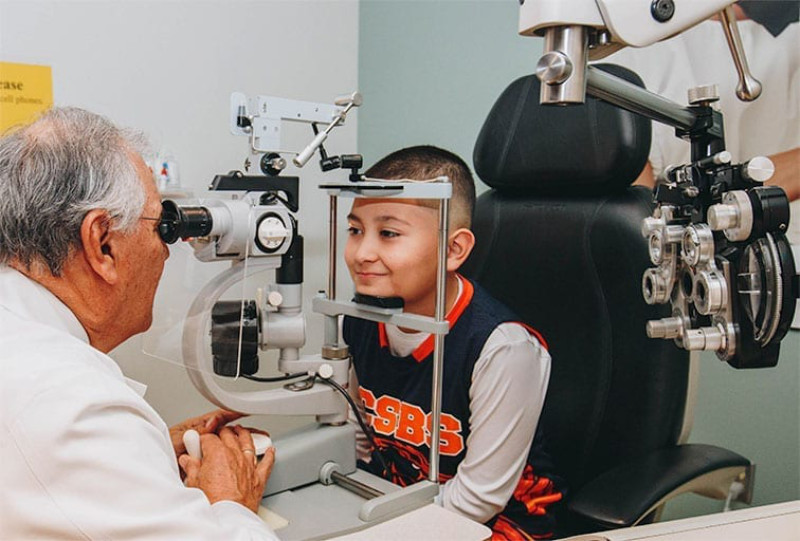
Yearly eye examinations are recommended to help detect and treat eye conditions in a timely manner. Routine eye examinations include several tests that directly or indirectly evaluate vision and eye health. Best-corrected vision is determined by having a person read letters, words, numbers or shapes. A person’s vision may be improved with the help of corrective lenses. The doctor through a procedure called refraction determines this. Eye alignment and eye muscle function are monitored. Pupil response to light and peripheral vision testing help assess proper eye and brain Routine Eye Exams communication. Measure of focusing ability and the ability to converge or pull the eyes together helps detect problems that may affect reading comfort. By using a microscope and ophthalmoscope the doctor monitors eye health. The eyelids and lashes, front surface of the eye, structures within the eye including the iris, lens, retina, blood vessels and optic nerve head are all observed and studied to evaluate eye health. Eye health evaluation is aimed at detecting a wide range of abnormalities. Conditions of concern to most patients include the need for glasses, the presence of cataracts, diabetic retinopathy, macular degeneration and glaucoma. A comprehensive yearly exam is offered to all patients and is also geared to the patient’s individual needs.
Diabetic Eye Exams
Diabetes is a disease causing blood sugar to become elevated. Over time, elevated levels of blood sugar affect small blood vessels throughout organs in the body. Blood vessels within the eye are affected early in the disease process. Retinal changes that result from diabetes are termed diabetic retinopathy. Changes to the blood vessels include micro aneurysms, which are zones of thinning along the blood vessel walls that begin to form balloon-like out-pouching. Like a garden hose, the blood vessels begin to sprout leaks at the weaker zones. Because blood vessels are compromised, the retina may not receive an adequate blood supply. This may stimulate new blood vessel formation within the eye. These new blood vessels tend to grow away from the retinal lining of the eye and toward the center of the eye. These misdirected blood vessels can be pulled upon and may lead to bleeding or detachment of the retina. Eye examination is an excellent way of viewing blood vessels within the eye that are representative of the blood vessels found in other organs of the body.
A diabetic eye examination is much like a routine eye examination except that special attention is given to the retina when examining within the eye. Blood vessels overlying the retina are carefully inspected for micro aneurysms, small hemorrhages along side weakened blood vessels, and new blood vessel growth. Because of the importance of the retinal evaluation for a patient with diabetes, eye drops are instilled into the eye to help dilate or temporarily enlarge the pupil. The pupil is the window into the eye. By enlarging the window, a better view within the eye is made possible.
Results of the eye examination can be relayed to the patient’s primary care provider via our State of the Art electronic medical records system which is capable of immediately sending them a letter via electronic mail, before our patient leaves the office
Computer Assisted Vision Tests
Vision is assessed in different ways. The primary method of measuring vision incorporates single letters of known size at a set testing distance read aloud by the patient. Ideal test conditions include optimum contrast, symbols recognizable to the patient, and various targets to avoid patient memorization. The typical letter chart used by most eye examiners uses a light projector to display black letters against a lighted background in a dark room to provide best contrast.
At Gulf Coast Eye Institute, the latest technology in vision testing is incorporated into the eye exam. A computer-based vision test provides more features than the traditional slide projector. With the use of a computer monitor as a direct lighting source, ideal contrast between a black letter, or figure and a white background can be achieved even in a well-lighted room. Optimum contrast is always available when measuring vision. Contrast can also be reduced to assess what is called contrast sensitivity, a different measure to assess vision. The use of various shades of gray letters against a white background is available at the click of a button. Such measures can be helpful in detecting vision problems that often escape detection with high contrast testing. The computer chart also offers very crisp letters through the use of a high-resolution computer screen. Various targets are available for identification to serve a variety of patient populations. Traditional letter charts such as the Snellen, Sloan, and ETDRS, can be selected for use as well a variety of other letter-based letter charts such as the Tumbling E, HOTV, and Landolt C. Every line size for every chart can be randomized or rearranged at the click of a button to help prevent patient memorization and more accurately measure vision. Symbols other than letters are available such as numbers or various shapes such as an apple, square, circle and house, recognizable to most preschool aged children. Such a range of options assists in accurately and repeatedly measuring vision in different ways.
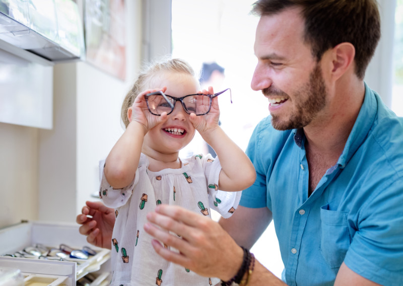
Pediatric Eye Exams
The American Optometric Association (AOA) and the American Association of Ophthalmology (AAO) suggest that children receive their first eye examination by one year of age. The first two years of life are a critical period for eye development. Proper eye development depends on clear images being received by both eyes as well as equal attention given to each eye. Conditions such as high refractive errors, big differences in refractive errors between the two eyes, and eye misalignment can have long-term affects on a person. Fortunately, such conditions can easily be detected and treated if diagnosed at an early age. Eye problems such as congenital cataracts and retinal diseases, although uncommon can occur in young children. Early detection is always preferred over delayed detection as treatment is more successful with early detection.
Even though children may not be able to communicate verbally like their parents, many of the same tests used in an adult examination can be administered to infants and children. The only difference is that measurements are made objectively by examiner observation. Eye alignment, depth perception, and eye health can all be assessed. Even vision can be assessed through modified tests designed for nonverbal patients. Early and regular eye examination can help ensure the best vision and eye care for your loved ones.
Foreign Body Removal
An ocular foreign body is an unintended object present on or within the eye. A foreign body may be metallic or non-metallic. Non-metallic objects can be an eyelash, a piece of glass, dirt, plastic, wood, plant matter, saw dust, or pretty much anything. Such objects are usually detected because of ongoing patient discomfort and signs of eye irritation. An object may approach the eye slowly or at fast speed. The size, shape and speed of approach to the eye can determine if an object is superficial, penetrates the eye or double perforate an eye. Eye irritation following grinding activities or following the use of power tools raises concern for a penetrating foreign body. Signs of eye penetration may include an object entry site and damage along the object’s path, which may be subtle or obvious depending on the foreign body. If an object penetrates the eye, it may need to be removed surgically, or may remain depending on the consequences of removing the object versus leaving it in place. A magnetic metallic object within the eye requires patient education. If the magnetic metallic object remains in the eye, the individual must inform their medical doctors and any health care provider that may request magnetic resonance imaging tests (MRI). Such testing involves the use of powerful magnets that will move magnetic fragments causing further damage to the eye and near by structures. If an object is superficial it can removed using fluid pressure, or mechanical force with simple objects such as a q-tip. If the object is slightly embedded, it may be removed using magnification provided by a Biomicroscope (Slit Lamp) an ocular foreign body tool kit. The best treatment for ocular foreign bodies is prevention. Many times an ocular foreign body can be avoided by exercising good judgment. Wearing approved safety goggles to protect the eyes when indicated can often prevent the need for ocular foreign body removal by an eye doctor.
Specialty Contact Fitting
Contact lenses certainly don’t fall under the one-size-fits-all umbrella. You might even have been told contacts aren’t ideal for you because you have “hard to fit” eyes. No worries.
The team at Gulf Coast Eye Institute helps many “hard to fit” patients find the ideal contact lens solution for their unique eyes.
Gulf Coast Eye Institute is pleased to offer SynergEyes lenses. SynergEyes makes hybrid contact lenses for patients with astigmatism, presbyopia and irregular corneal conditions. If this is you, great news! You can now see life with crystal clear vision, clarity and comfort all day long. This is the only hybrid lens that is highly breathable, made from healthy materials and is specifically designed for people with irregular corneas.
Amniotic Membrane Therapy
Advances in medical research mean there are now some amazing treatment options for damage and scarring on the surface of the eye. Using an amniotic membrane graft (basically a bandage made of biologically correct tissue instead of artificial ingredients), the doctors at Gulf Coast Eye Institute can implant amniotic membrane to encourage the healing process in eyes that have been affected by keratitis, corneal ulcers, neurotrophic keratopathy, chemical burns or even severe dry eye. The procedure is non-invasive and non-surgical; the membrane is placed on a contact lens-like structure that is gently placed on the eye. The membrane slowly dissolves behind the contact lens to heal the surface of your eye, and the contact is later removed.
If amniotic membrane therapy could help your healing process, your Gulf Coast Eye Institute ophthalmologist will answer any questions you have when discussing the treatment.